
Figure 3.
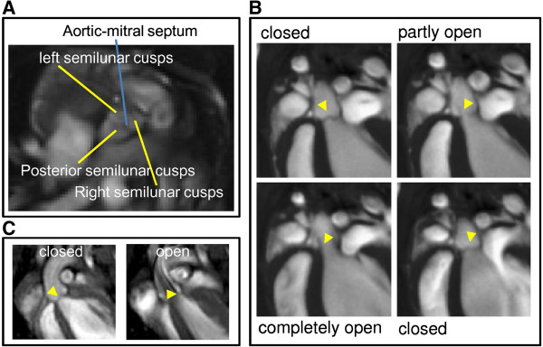
Self-gated cine FLASH and UTE images of the aortic valve. Self-gated UTE images (A) in short-axis orientation visualizing the aortic-mitral septum and (B) long-axis view of the aortic valve (yellow arrowhead) in different states during the heart cycle. (C) Self-gated FLASH images of the aortic valve (yellow arrowhead) in closed and open position.