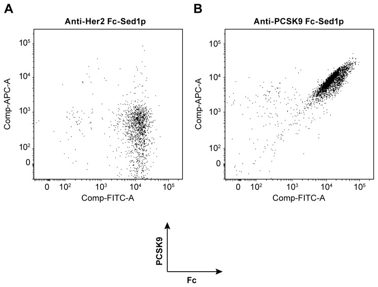
Figure 3. Antigen binding capacity of monovalent IgGs displayed by Fc-Sed1p measured by flow cytometry.
The cells were dually labeled with goat anti-human Fc DyeLight 488, biotinylated PCSK9, and APC 635 labeled Streptavidin A) FACS analysis of labeled Pichia pastoris strains displaying Fc-Sed1p complexed with monovalent anti-Her2 antibody fragment (H+L) or B) an monovalent anti-PCSK9 (H+L) antibody fragment.

