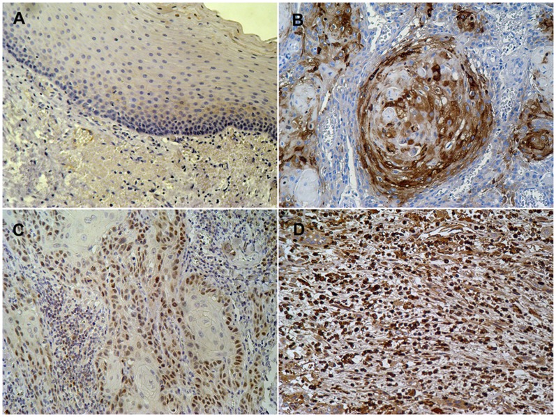
Figure 1. Expression patterns of Snail in OSCC specimens.
(A) Very faint immunoreactivity of Snail was observed in normal epithelial specimens (200X). (B) In well-differentiated oral squamous cell carcinomas (OSCCs), low immunoreactivity expression surrounding the epithelial pearl, while centrally situated cells remained weakly positive (200X). (C) In moderately differentiated OSCCs, homogeneous and moderate staining for Snail was labeled in diffuse type at the peripheral of the nest (200X). (D) In poorly differentiated OSCCs, individual tumor cells were observed for Snail labeling and also had a stronger Snail staining (200X).

