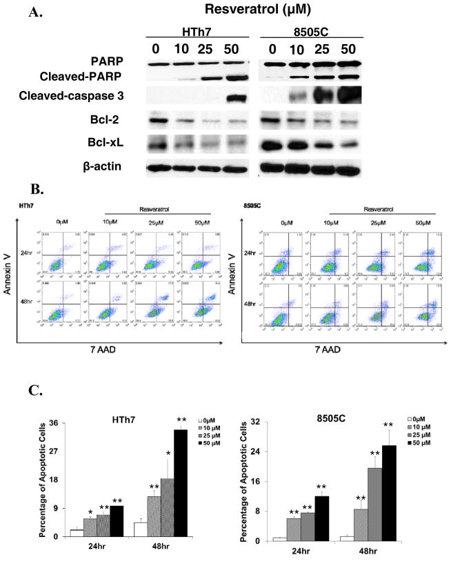
Figure 3.
Apoptosis in ATC induced by resveratrol treatment. A, Detection of apoptotic markers including cleaved-PARP, cleaved-caspase 3, Bcl2 and Bcl-xL by Western blot in HTh7 and 8505C cells treated with three different concentrations of resveratrol (10, 25, 50μM) or vehicle control. Equal loading was confirmed with β-actin. B, Resveratrol induced apoptosis in a time-and dose-dependent manner. ATC cells were exposed to different concentrations of resveratrol for 24 hours and 48 hours before they were double stained with Annexin V and 7 AAD for flow cytometry analysis. Percentages at right lower quadrant denote the cells in early apoptotic phase. Results of one representative experiment. Data from three repeated experiments are summarized in bar graph (C), and are shown as mean ± SD (n=3, *P<0.05, **P<0.01 vs. control cells without resveratrol treatment).

