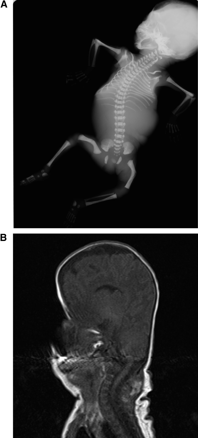
Figure 1.
A, An x-ray radiograph performed at postmortem after the termination of pregnancy PORD B at gestational week 25; note the abnormally shaped skull, thin ribs, ankylosis of both elbow joints (radioulnar synostosis), and bilateral bowed femora. B, Magnetic resonance imaging of the head of patient PORD B on postnatal day 2, illustrating the pronounced turricephaly consequent to PORD-associated craniosynostosis.

