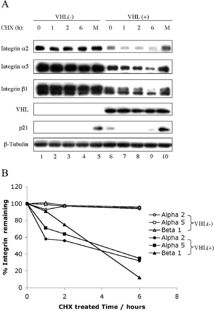
Fig. 3.
VHL leads to increased proteasome-dependent degradation of integrins. (A) Confluent VHL-negative 786-0 cells containing pCR3 and VHL-positive 786-0 cells ectopically expressing VHLp18(MEA) were treated with cycloheximide (CHX) for the times indicated about each blot or with both CHX and MG132 for 6 h. Cell lysates were separated in a 4~20% SDS-PAGE gel and immunoblotted with antibodies indicated to the left of the blots. Integrin α3 is undetectable in 786 (VHL+) cells (see Fig. 1). p21, a short-lived proteasomally degraded protein, was used as a positive control for CHX/MG132 treatment. (B) Integrin band intensities were quantified by IMAGEUANT 5.0 (Molecular Dynamics, Sunnyvale, CA) and are graphically represented.

