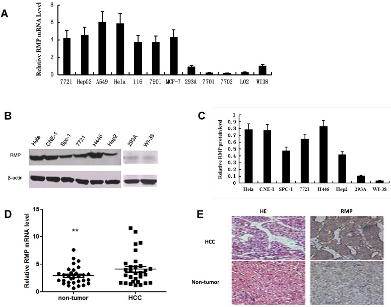
Fig 1.
RMP expression is increased in cancer cells and HCC tissues. (A) The mRNA expression of RMP in normal and cancer cell lines was examined by qRT-PCR as described in Materials and Methods. The relative mRNA levels of RMP was normalized against GAPDH and depicted graphically. (B) The RMP protein expression was examined by Western blot analysis as described in Materials and Methods. The relative levels of RMP protein expression was normalized against β-actin and depicted graphically in (C). Results are reported as mean +/-S.D. of three independent experiments. (D) Higher expression of RMP in HCC. Total RNA was extracted from 30 pairs of tumor and matched non-tumor tissues of HCC patients. Quantitative real-time RT-PCR was performed for the detection of RMP mRNA. The expression levels RMP mRNA were shown in close dots (Non-tumor) and squares (HCC). The relative expression levels of RMP mRNA was normalized against β-actin. Student's t test, **, p<0.01, relative to controls. (E) The tumor and matched non-tumor tissues from HCC patients were sectioned and subjected to H-E staining and immunostaining with antibody against RMP. The sections were observed in a microscope at 400 magnifications. Cell line abbreviation: 7721 and HepG2: Human Hepatocellular carcinoma cells; A-549: Human lung adenocarcinoma A-549 cells; HeLa: Human cervical carcinoma HeLa cells; 116: Human colon carcinoma HCT-116 cells; 7901: Human gastric carcinoma SGC-7901 cells; MCF-7: Human breast carcinoma MCF-7 cells; 293A: Normal human kidney QBI-293A cells; WI-38: Normal human diploid embryonic lung fibroblasts WI-38 cells; 7701, 7702 and L02: Normal hepatic cell lines of QSG-7701, HL-7702 and L02; CNE-1: Human nasopharyngeal carcinoma CNE-1 cells; Spc-1: Human lung adenocarcinoma Spc-1 cells; H446: Human small cell lung cancer H446 cells; Hep2: Human caucasian larynx carcinoma squamous Hep2 cells.

