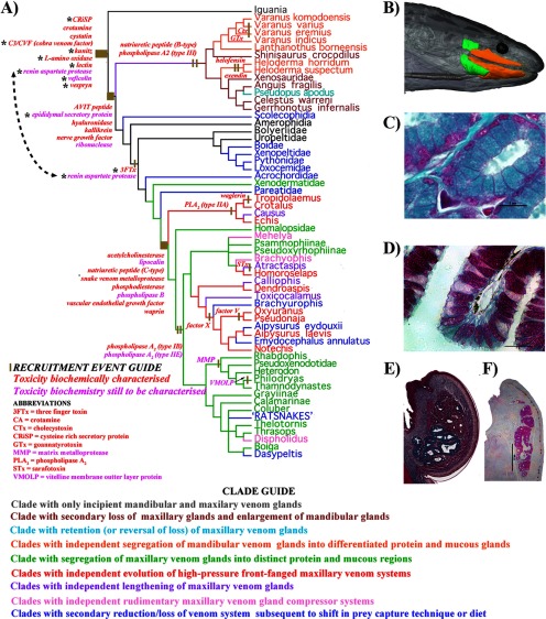
Fig. 1.
Cladogram of evolutionary relationships of toxicoferan reptiles (Fry et al., 2006; Lee, 2009; Piskurek et al., 2006; Vidal and Hedges 2002, 2004, 2005, 2009) showing relative timing of toxin recruitment events and derivations of the venom system. A, * indicates toxin type with relative gene recruitment event timing changed because of results from this study. B, Magnetic resonance imaging of C. ruffus showing the rictal (green) and maxillary/mandibular glands (orange). Histochemically stained in B Masson's Trichrome C. ruffus rictal gland and C, Periodic Acid Schiff's Aspidites melanocephalus mandibular gland; both with 5 μm histology scale bars. Mandibular glands stained with D Masson's Trichrome Anolis equestris showing the mixed sero-mucous gland or E Periodic Acid Schiff's Lepidophyma flavimaculatum showing the purely mucous secreting gland. n = nucleus; S.E. = secretory epithelial cells (S.E.); V = enlarged vesicles.

