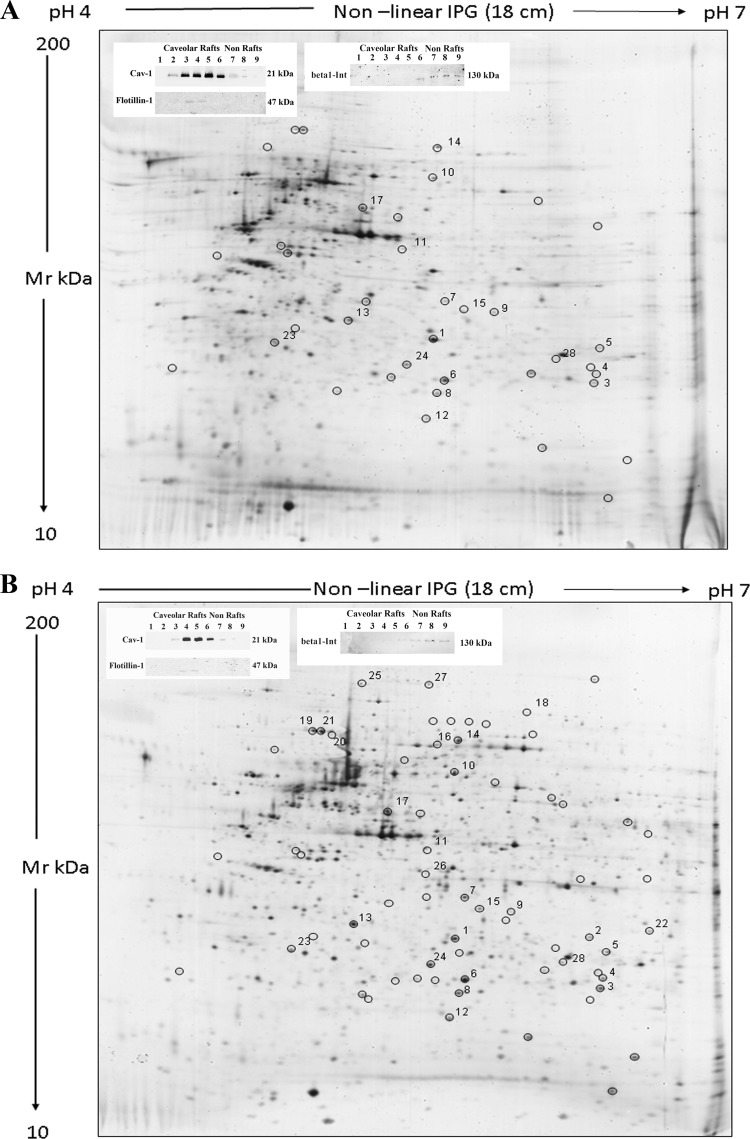
Fig. 1.
2-D gels of caveolar-raft fractions. Silver-stained 2-D gels of caveolar-raft membrane fractions from control (A) and VEGF-stimulated ECFCs (VEGF: 50 ng/ml for 12 h) (B). Proteins (40 μg) were separated by their pI on 4–7 linear IPG strips and by their molecular weight on 9–16% polyacrylamide linear gradient gels. Black circles indicate spots that presented a differential occurrence in micro-domain after VEGF stimulation. Numbers indicate spot identified by mass spectrometry. The insets of each panel show the Western blotting with anticaveolin, antiflotillin and anti-integrinβ1 antibodies of fractions obtained by density-gradient centrifugation of ECFCs lysates (the caveolin-rich fractions Western blotting in each panel is representative of 20 different experiments performed with preparative purposes, to collect the amount of material to be subjected to mass-spectrometry analysis). Caveolin and flotillin positive fractions were collected and used in the 2-D gels. Molecular weights are reported on the right.

