
Fig. 2.
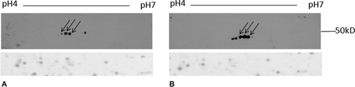
uPAR detection by Western blotting. Caveolar-raft proteins (150 μg) were separated by their pI on 4–7 linear IPG strips and by their molecular weight on 9–16% polyacrylamide linear gradient gels. After 2-DE, gels were blotted on PVDF membrane and uPAR was detected with specific antibody. Arrows represent corresponding spots between the two blots. A, control cells; B, VEGF stimulated cells. The same blots were stained with Coomassie brilliant blue (a representative part of PVDF membrane is reported) to confirm that protein load was the same.