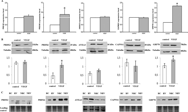
Fig. 3.
Quantitative RT-PCR and Western blotting analisys of selected caveolar raft-enriched proteins before and after VEGF stimulation. A, Real time PCR showing gene expression levels of calpain small subunit-1 (CAPNS1), peroxiredoxin-2 (PRDX2), peroxiredoxin-6 (PRDX6), annexin-A3 (ANXA3), and 78 kDa glucose-regulated protein (GRP78). Transcripts were quantified by real-time PCR in ECFCs before and after 4 h of VEGF challenge (50 ng/ml). Data shown are the mean of three different experiments performed in triplicate. The two-tailed, nonpaired Student's t test was performed. * indicates a p value < 0.05. B; Western blotting analisys of selected proteins in whole lysates before and after 12 h VEGF treatment (50 ng/ml). Intensity of immunostained bands was normalized against tubulin. Histograms, reporting the normalization in arbitrary units, are the mean of three different experiments performed in triplicate. The two-tailed, nonpaired Student's t test was performed. * indicates a p value < 0.05. C, Western blotting analysis of selected proteins in caveolar-raft and nonraft fractions before and after 12 h of VEGF treatment. The protein load was checked by staining the PVDF membrane with Coomassie. RC: raft fraction, control; RV: raft fraction, VEGF stimulation; NRC: non-raft fraction, control; NRV: non-raft fraction, VEGF stimulation. Each blotting shows the result of a typical experiment of three that gave similar results.

