
Figure 10.
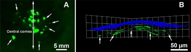
Representative stereomicroscopy (A) and confocal microscopy (B) images showing transgene delivery in the rabbit stroma in vivo noted 2 days after topical application of transfection solution (1 μg/μl plasmid in 50 nmol DDAB and 50 nmol DOPE in 100 μl lactated Ringer's solution) onto the rabbit cornea via custom delivery technique. The plasmid expresses transgene under control of CMV+chicken-β-actin promoter. Nuclei are stained blue with DAPI.