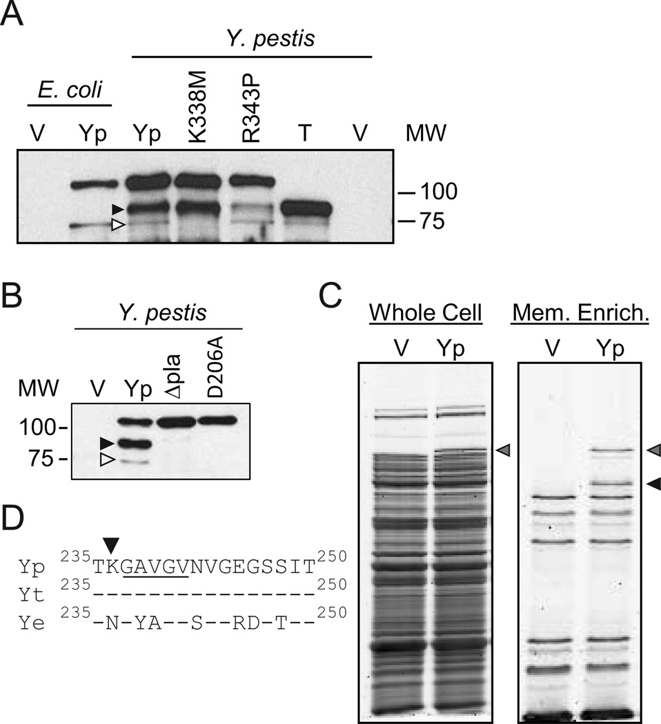
Figure 4. Identification of Pla cleavage sites in YapE.
(A)To identify the Pla cleavage sites in Y. pestis YapE, YapE point mutants that altered OmpT cleavage of YapE were expressed in Y. pestis and Western blots were performed with anti-YapE serum. V = Vector only (pMWO-034); Yp = WT Y. pestis YapE (pMBL313); K338M = K338M pt. mutant (pLOU021); R343P = R343P pt. mutant (pLOU022); T = YapEΔ34-236 (pLOU109). As a reference, V and Yp samples expressed in E. coli are also included. (B) Anti-YapE Western blots of Y. pestis YapE expressed in Y. pestis (Yp), Y. pestis Δpla, or Y. pestis with catalytically inactive Pla (D206A). V = vector control. (C)Whole cell proteins and outer membrane enriched samples from Y. pestis stained with Flamingo fluorescent stain. Yp = Y. pestis YapE (pMBL313); V = Vector control (pMWO-034). (D) Alignment of YapE proteins between amino acids 235 and 250. Yp = Y. pestis YapE, Yt = Y. pseudotuberculosis YapE; Ye = Y. enterocolitica YapE; underlined region represent the N-terminal amino acids of the Pla cleaved YapE peptide. Gray arrowheads represent full length YapE, white arrowheads represent YapE OmpT cleavage site/product, and black arrowheads represent primary YapE Pla cleavage site/product.

