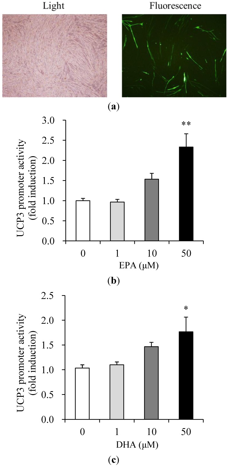
Figure 3.
Effects of EPA and DHA on UCP3 promoter activity in muscle cells. Transfection efficiency (a) of differentiated C2C12 muscle cells was analyzed with a GFP expression vector. The GFP expression was observed under fluorescent microscope at 40× magnification. Cells were transfected with the UCP3 (−1790/+52 bp)/luc reporter gene and pCMV-β galactosidase, and were then incubated 1% BSA serum-free medium with the concentrations of EPA (b) and DHA (c) indicated, from 0 (control) to 50 μM, for 40 h. Promoter activities measured by luciferase activity were calculated in relative light units (RLU) and normalized to β-galactosidase activity. Values are expressed as mean ± SE (n = 3) of three independent experiments. * P < 0.05 and ** P < 0.01 versus control treatment.

