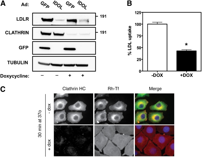
Fig. 2.
IDOL-mediated degradation of the LDLR is clathrin independent. A: Inducible CHC knockdown HeLa cells were cultured in the presence (+) or absence (−) of doxycycline for 6 days to induce silencing of the CHC. Subsequently, cells were infected with a GFP- or IDOL-expressing adenovirus for 10 h at an MOI of 5. Total cell lysates were immunoblotted as indicated. B: Inducible CHC knockdown cells were cultured as in (A). Subsequently, cells were shifted to sterol-depletion medium for 16 h and then incubated for 30 min with 5 μg/ml DyLight 488-labeled LDL. After extensive washing, internalized LDL was quantified by measuring fluorescence in total cell lysates. LDL uptake in −Dox cells was set to 100%. Each bar and error represent the average ± SD (n = 8; *P < 0.01; DOX/dox, doxycycline). C: Inducible CHC knockdown cells were cultured as in (A) and then treated with rhodamine-transferrin (red) for 30 min. After fixation, cells were stained for clathrin (green), and counterstained with DAPI (blue). Note that in clathrin knockdown cells transferrin internalization is blocked, while in control cells transferrin is internalized and colocalized with clathrin. Immunoblots are representative of three independent experiments.

