
Figure 3.
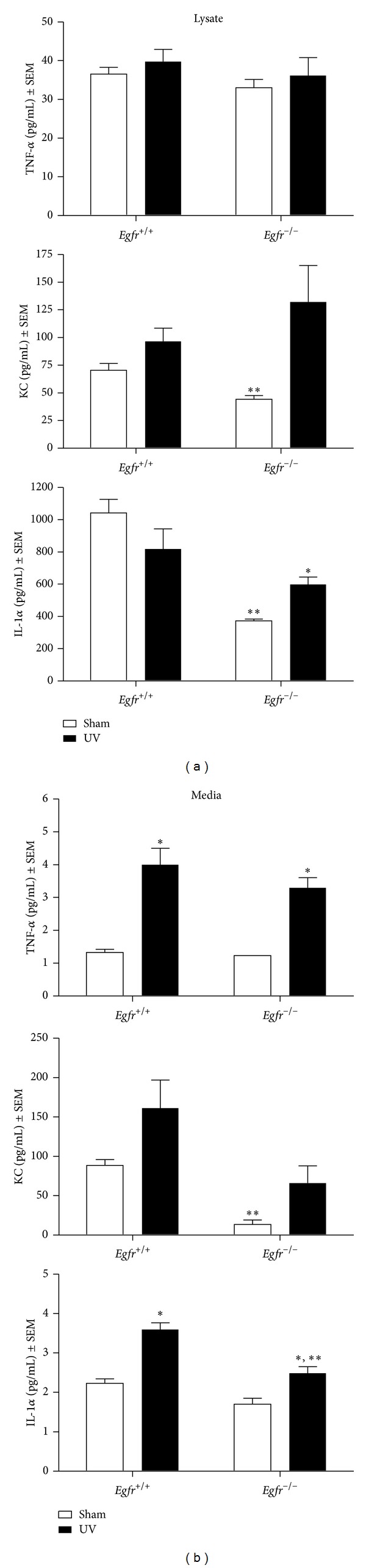
Genetic deletion of Egfr reduces TNF-α, KC, and IL-1α in keratinocytes following UV exposure. Subconfluent Egfr-null and wild-type keratinocytes were exposed to 600 J/m2 UV irradiation and cell lysate (left panel) and media (right panel) were harvested for cytokine analysis at 16 h using a Luminex instrument. N ≥ 4 dishes. *Mean is significantly different from the corresponding sham-irradiated control or **significantly different from the corresponding wild-type group, using a Student's t-test, where P ≤ 0.05.