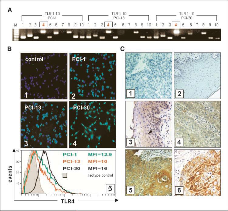
Figure 1.
TLR expression in tumors. TLR expression at the mRNA and protein levels in tumor cell lines, tumor, or normal mucosa. A, expression of TLR1 to TLR10 mRNA in the three HNSCC cell lines. B, expression of TLR4 protein in the three HNSCC cell lines. Magnification, ×400. 1, negative staining for TLR4 using isotype control IgG; 2, TLR4 in PCI-1; 3, TLR4 in PCI-13; 4, TLR4 in PCI-30 cells; 5, mean fluorescence intensity of TLR4 as determined by flow cytometry in the three cell lines. C, immunohistochemistry for TLR4 in tissue sections. Magnification, ×400. 1, isotype control in the tumor; 2, isotype control in the normal mucosa; 3, several TLR4+ cells in the normal mucosa (arrow); 4, a poorly differentiated HNSCC (G3) showing a weak positive reaction for TLR4; 5, a well-differentiated HNSCC (G1) showing strong positive reaction; 6, a representative HNSCC stained for cytokeratin.

