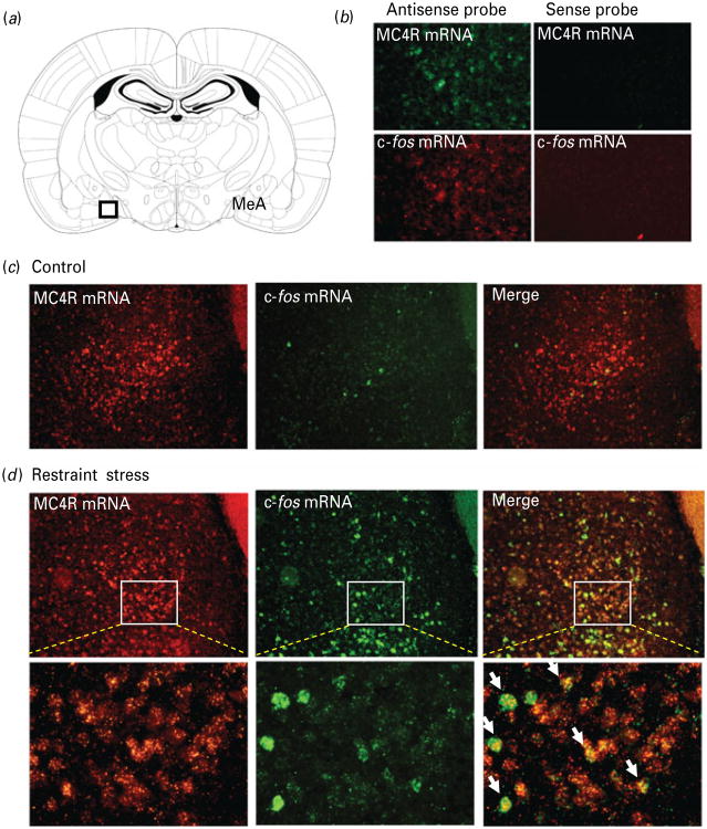
Fig. 1. Acute restraint stress activates melanocortin-4 receptor (MC4R)-expressing neurons in the medial amygdala.
Double-labelling fluorescent in situ hybridization showing the co-localization of c-fos mRNA (green) with MC4R mRNA (red) in the medial amygdala (MeA). (a) Schematic diagram of coronal brain section through the amygdala (Paxinos & Watson, 1998); (b) double-labelling fluorescent in situ hybridization using sense and anti-sense cRNA probes to detect expression of MC4R and c-fos mRNA in rat MeA; (c) representative images showing c-fos mRNA and MC4R mRNA expression in the MeA from a naive rat; (d) representative images showing the co-localization of c-fos mRNA and MC4R mRNA expression in the MeA from a rat exposed to 30-min restraint stress. The white arrowheads indicate cells double-labelled for c-fos mRNA and MC4R mRNA.

