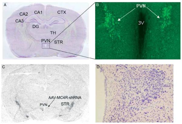
Figure 6.
AAV-mediated MC4R knockdown in the paraventricular nucleus of the hypothalamus. AAV-control-shRNA and AAV-MC4R-shRNA vectors were stereotaxically delivered into one side of the PVN of the rat. The animal was killed at 35 days after microinjection of AAV vectors. (A) Nissl-stained coronal brain section. (B) Fluorescent microscopic image showing EGFP expression, indicating the site of injection of AAV vectors. (C) In situ hybridization autoradiogram showing suppression of MC4R mRNA expression on the side of the PVN infected with AAV-MC4R-shRNA vectors. (D) Bright-field microscopic image showing no necrosis or abnormal cytoarchitecture in the PVN. CA1–3, area 1–3 Ammon’s horn; CTX, cortex; DG, dentate gyrus; PVN, paraventricular nucleus of hypothalamus; STR, striatum; TH, thalamus.

