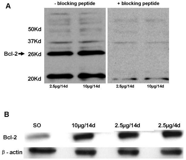
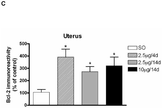
Figure 2.
Comparison of levels of Bcl-2 protein in uterus following various E regimens on Western blots. (A) Western blots of uterine extracts demonstrating the specificity of the antibody to Bcl-2. Bcl-2 antibody was preincubated with the blocking peptide Bcl-2P as described in Materials and Methods. Blots of uterine extracts from animals treated with 2.5 μg and 10 μg E for 14 days were treated with the antibody-antigen solution displayed no immunolabeling for the Bcl-2 band at 26 kDa. (B) Representative Western blot of Bcl-2 and β-actin in whole uterine extracts from animals treated with the various E regimens. Proteins (20μg) were resolved on SDS-PAGE (12.5%) and were then transferred to polyvinylidene fluoride transfer membranes using electrophoresis. Detection and quantification of proteins were performed as described in Materials and Methods. All E treatments resulted in an increase in Bcl-2 immunolabeling relative to SO. (C) Effects of various E regimens on Bcl-2 protein levels in whole uterine extracts. E treatment groups are significantly different from the control group SO (U values=2, 2, 0 respectively; P<0.01). Values are mean ± S.E.M, and are represented as the percentage of control. *: P<0.01, compared with SO.


