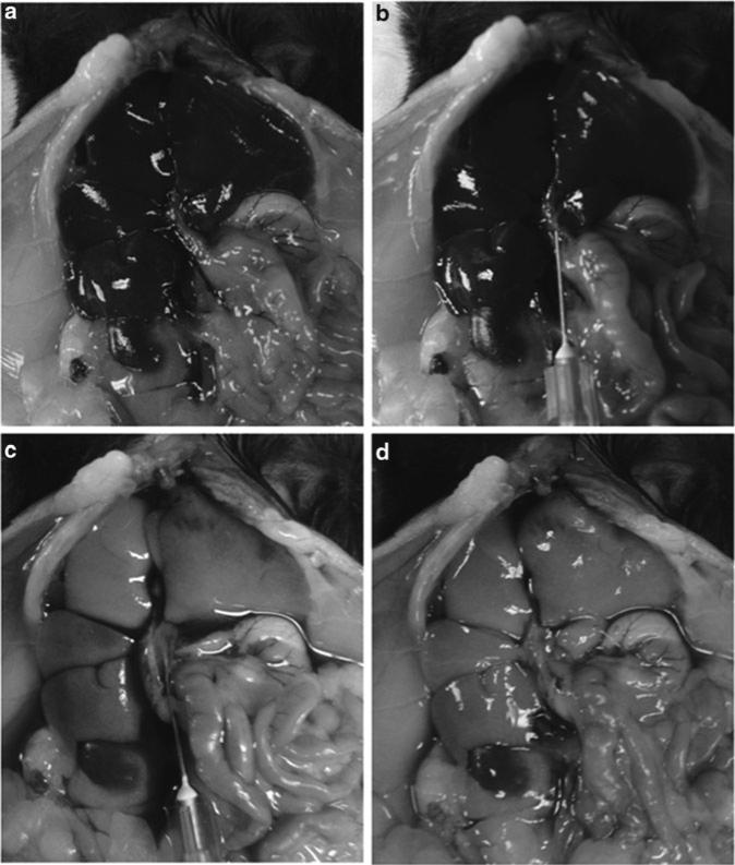
Fig. 3.
Perfusion of a mouse liver. (a) Situation before perfusion with portal (right) and inferior cava (left) vein entering the liver. (b) Insertion of a needle into the portal vein. (c) Start of perfusion of the liver. (d) Situation after perfusion of the liver. Please note the change in liver color after perfusion as a sign of successful removal of blood cells.

