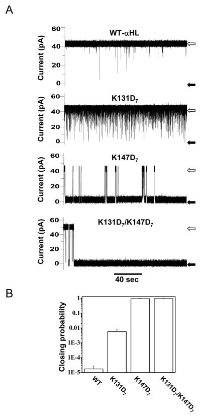
Fig. 4.
Different single-channel electrical signatures given by the interactions of the pb2(95)-Ba protein with various engineered αHL protein pores. (A) single-channel electrical recordings shows that pb2(95)-Baproteins interact more strongely with the pore (channel almost closed, black arrows) when the two electrostatic traps are present in the pore. The wild-type αHL protein pore shows the weakest interaction (channel mostly open, white arrows), as also indicated by the residence probability; (B) The residence probability is obtained by adding the channel closure times and dividing by the total recording time. 200 nM pb2(95)-Ba was added to the trans chamber. Single-channel recordings were performed as in Fig. 3C. Figure was reproduced from Ref. (27).

