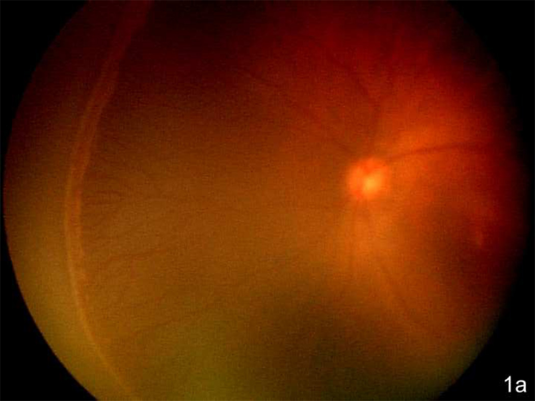
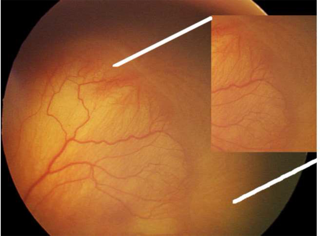
Figure 1.
a. Zone II with ridge in the nasal periphery of the left eye of a preterm infant showing early stage 3 ROP, or intravitreous neovascularzation (Retcam image, Clarity, CA).
b. Left eye of preterm infant with stage 3 ROP (intravitreous neovascularization) showing flat neovascularization that can occur in zone I and posterior zone II ROP. (see inset zoomed image; Retcam image, Clarity, CA).


