Figure 5.

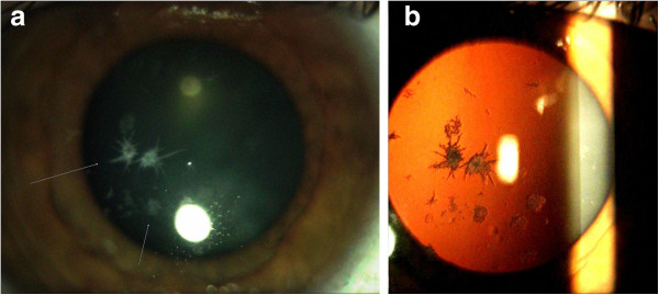
Slit lamp photography in the left eye (Case 2). The arrow indicates intense white snowflake-like deposits and rare gray-white granular deposits in mid-stromal layer (Panel a). Retroillumination of corneal deposits (Panel b).

Slit lamp photography in the left eye (Case 2). The arrow indicates intense white snowflake-like deposits and rare gray-white granular deposits in mid-stromal layer (Panel a). Retroillumination of corneal deposits (Panel b).