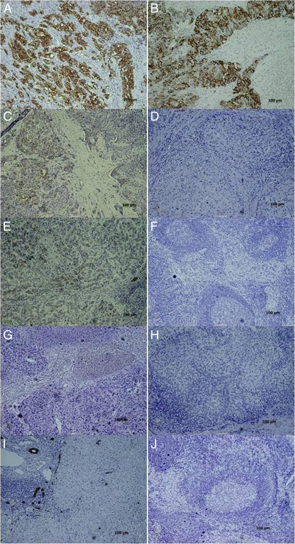
Figure 4.

Immunohistochemical microphotograph of primary liver tumor (left column: A, C, E, G, and I) and regional lymph nodes (LN) (right column: B, D, F, H, and J) for CK19 expression. A and B) CK19 (+) primary liver tumor with CK19 (+) lymph node metastasis (LNM). The cytoplasmic staining of CK19 can be demonstrated in both primary tumor and metastatic LN. C and D) CK19 (+) primary liver tumor with CK19(−) LNM. The regional lymph nodes had been infiltrated by metastatic tumor cells, which did not express CK19. E and F) CK19 (+) primary liver tumor without LNM. The normal LN structure can be clearly identified. G and H) CK19 (−) primary liver tumor with CK19(−) LNM. Both the primary tumor and metastatic LN did not express CK19 in their cytoplasm. On the other hand, the biliary epithelial cells expressed CK19. I and J) CK19(−) primary liver tumor without LNM. (Magnifications, x100).
