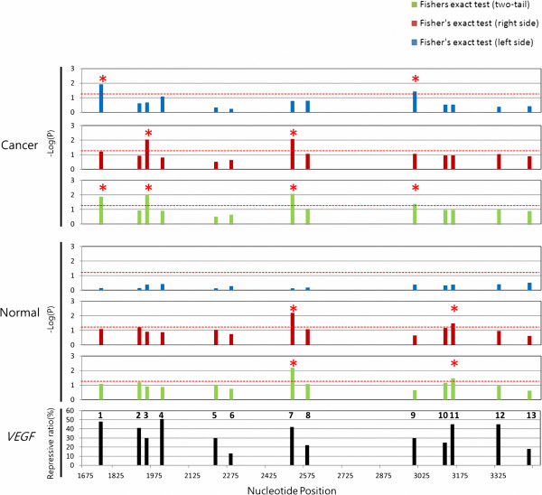
Figure 4.
Comparison of splicing-regulated MRE regions and highly repressive MREs in VEGF gene. For the alternative splicing analysis of the 13 MRE regions, the top, middle, and bottom panels present the results obtained for cancer tissues, the results obtained for normal tissues, and the repressive ratios reported in the literature, respectively. The negative log10-transformed P-values for the 13 MRE regions (numbered in the bottom panel) are presented in blue, red, and green and correspond to the left-side, right-side, and two-tail Fisher exact tests, respectively. The red star indicates statistical significance at P < 0.05, determined by Fisher exact test. Highly repressive MREs with repression ratios above 30% are found in MRE regions 1, 2, 3, 4, 5, 7, 9, 11 and 12. Putative hsa-miR-148 recognition sites in human DNMT3B coding region. Dotted red line indicates significance region (P < 0.05).

