
Figure 3. Loss of pericardial cell adhesion causes loss of heart function.
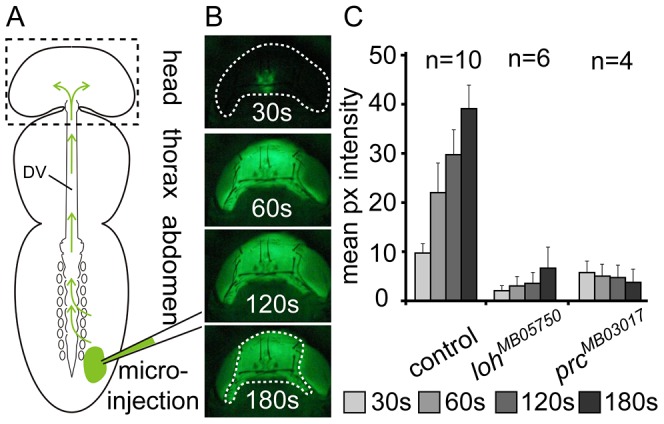
(A) Scheme depicting the basics of dye angiography in pharate adults. The main body parts, the dorsal vessel (DV) and the injection area are indicated. (B) Head of a wild type animal (corresponding to dashed box in scheme A) showing the accumulation of the tracer at four consecutive time points after injection. (C) Mean pixel intensities measured at four consecutive time points showing cardiac output in wild type (white1118) and homozygous prcMB03017 and lohMB05750 pharate adult animals. Error bars are s.e.m. The region used for measurement is indicated in the lowest panel in B.