
Figure 4. Molecular characterization of loh and prc.
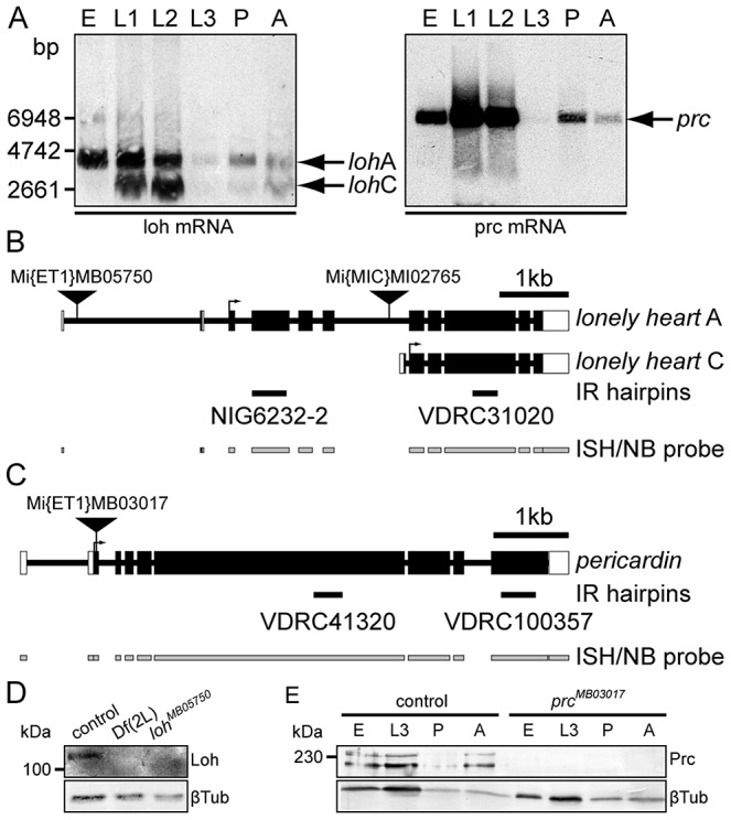
(A) Developmental Northern blots showing loh and prc expression in total RNA samples of 0–24 h old embryos (E), first, second or third instar larvae (L1–L3), mid-stage pupae (P) or adults (A) using gene specific riboprobes (indicated in B and C). (B, C) Schematic representation of loh (B) and prc (C) gene loci and transcripts. The schemes indicate the position of transposons, location of hairpins (IR) used for knock down and riboprobes used for Northern analysis (NB) and in situ hybridization (ISH). (D) Immunoblot of total protein extracts obtained from stage 17 control, homozygous Df(2L)Exel7048 or homozygous lohMB05750 embryos probed with antibodies against Loh or βTub. Loh is undetectable in homozygous deficiency or mutant extracts. (E) Immunoblot of total protein extracts obtained from control or homozygous prcMB03017 0–24 h old embryos (E), third instar larvae (L3) mid-stage pupae (P) or adults (A) probed with antibodies against Prc or βTub. Prc is undetectable in extracts of homozygous mutants.