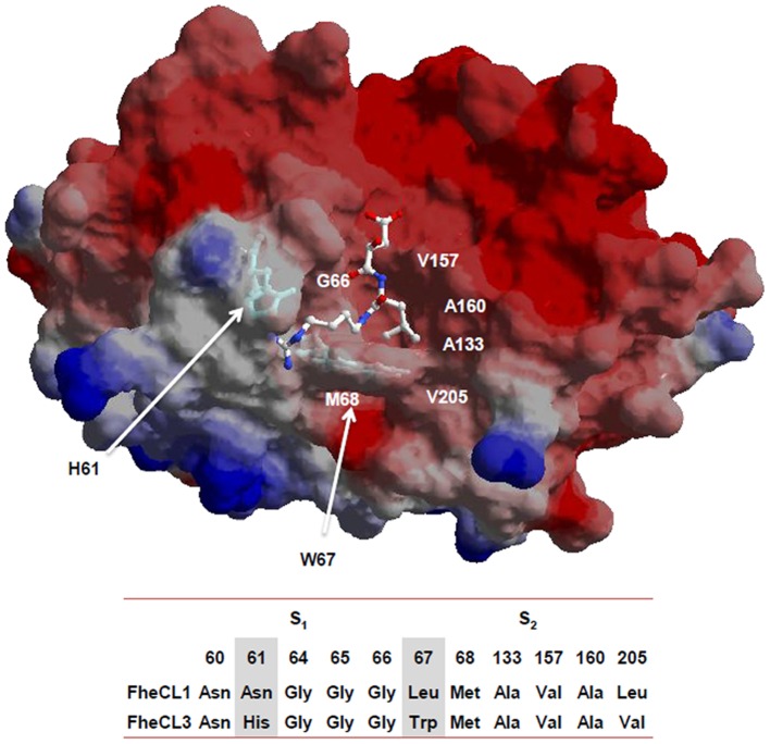
Figure 4. Residues contributing to substrate binding.
Model of mature FheCL3 showing the S2 and S3 subsites of the active site: the His61 and Trp67 residues that were mutated in the present study are highlighted as sticks. The E64 inhibitor complexed with human cathepsin K (1ATK) was superimposed to facilitate viewing the active site cleft. The image was generated wth SPDBviewer [33] The residues constituting the active site S2 and S3 subsites of FheCL1 and FheCL3 are indicated below the molecular representation.

