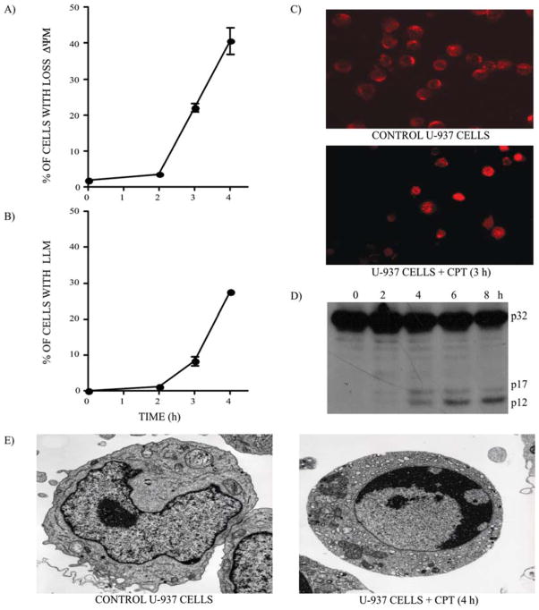
Figure 1.
CPT-induced apoptosis in U-937 cells. (A) ↓ΔΨm and (B) LLM were monitored in U-937 cells after CPT (1 μM) treatment. Bars represent the means of 4 independent determinations and error bars represent SEM. (C) Control and CPT-treated U-937 cells were stained with a specific antibody against cathepsin D to monitor the de-localization of lysosomal cathepsin D, 3 h after CPT treatment (1 μM). (D) Western blotting of caspase-3. The active cleaved 17 and 12 kDa fragments of procaspase-3 (32 kDa) are clearly visible 4 h after CPT treatment (1 μM). (E) Electron microscopy representative of the control and CPT-treated U-937 cells (4 h).

