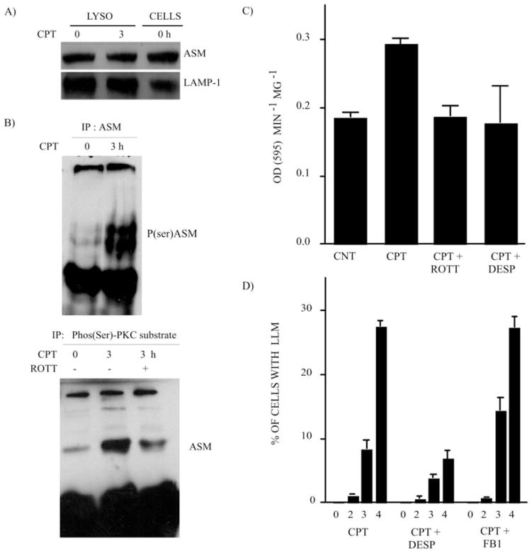
Figure 3.
PKC-δ mediates ASM phosphorylation and activation in lysosomes after CPT treatment. (A) ASM expression in highly-enriched lysosomal extracts from the control (CNT) and CPT-treated U-937 cells (1 μM; 3 h). Whole-cell extract (CNT) is shown as the ASM antibody control. LAMP-1 expression is the loading control. (B) Western blotting revealed the ASM phosphorylation level after CPT-treatment (1 μM; 3 h). IP was performed from highly-enriched lysosomal preparations. Upper panel, IP was undertaken with anti-ASM antibodies and Western blotting was carried out with anti-phospho-(Ser) PKC substrate antibodies. The doublet represents some protein degradation. Lower panel, reciprocal experiment where IP was performed with anti-phospho-(Ser) PKC substrate antibodies and Western blotting was performed with anti-ASM. (C) ASM activity monitored in highly-enriched lysosomal extracts from CNT and CPT-treated U-937 cells (1 μM; 3 h) in the absence and presence of the PKC-δ inhibitor, ROTT (3.5 μM), and the ASM inhibitor, DESP (10 μM). Bars represent the means of 3 independent determinations and error bars represent SEM. (D) LLM was monitored after CPT treament in U-937 cells where ASM activity was inhibited by DESP (10 μM), and CS activity was inhibited by FB1 (10 μM). Bars represent the means of 3 independent determinations and error bars represent SEM.

