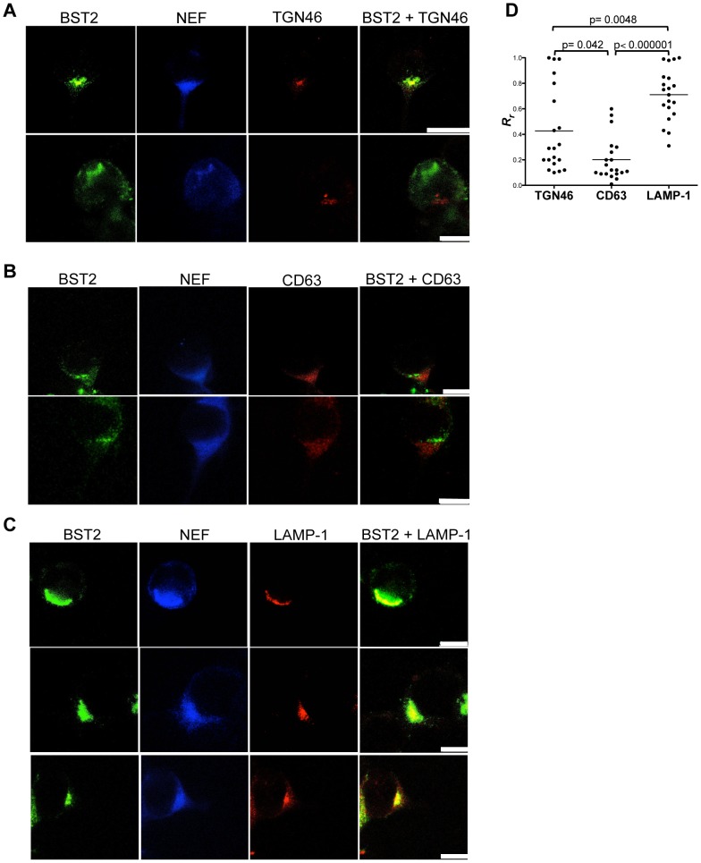
Figure 9. Subcellular distribution of tetherin in SIV-infected cells.
293T cells expressing HA-tagged rhesus macaque tetherin were infected with VSV-G pseudotyped SIVmac239 Δenv and stained for tetherin (green), Nef (blue) and either TGN46 (red) (A), CD63 (red) (B) or LAMP-1 (red) (C). The white scale bar indicates 10 µm. (D) The extent of co-localization between tetherin and each of the intracellular markers was estimated by calculation of the Pearson's correlation coefficients for images of twenty randomly selected SIV-infected cells.

