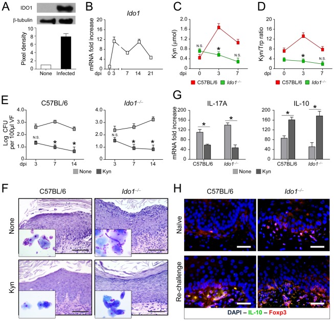
Figure 4. IDO1 and kynurenines mediate tolerance in murine VVC.
(A) IDO1 protein and (B) gene expression in the vagina of C57BL/6 mice (n = 4) intravaginally infected with C. albicans. Proteins in vaginal cell lysates (3 dpi) were visualized by western blotting with rabbit polyclonal IDO1 specific antibody. Scanning densitometry was done on a Scion Image apparatus. Western blots out of 2 independent experiments and corresponding pixel density ratio normalized against β-tubulin. Ido1 mRNA expression [normalized to mRNA of naïve (dpi 0) mice] in vaginal tissue (RT-PCR) at different dpi. (C) Relative concentrations of kynurenines (Kyn) and (D) kynurenine-to-tryptophan (Kyn/Trp) ratio in vaginal fluids at different dpi. Pooled results from 3 different experiments. *P<0.05, IDO1-deficient vs. C57BL/6 mice at the days indicated. N.S., not significant. (E) Vaginal fungal growth (Log10 CFU/100 µl VF ± s.e.m.) at different dpi in mice (n = 6) treated intraperitoneally with a mixture of l-kynurenine, 3-hydroxykynurenine and 3-hydroxyanthranilic acid or PBS (None). Pooled data from 3 different experiments.*P<0.05, treated vs. untreated mice at the days indicated. N.S., not significant. (F) Periodic acid-Schiff-stained vaginal sections and inflammatory cell recruitment in vaginal fluids (May–Grünwald Giemsa staining in the insets) acquired with a 40× and 100× objective, respectively, at 21 dpi. Scale bars, 100 µm. Representative image from 3 experiments. (G) Cytokine levels (pg/mg, cytokine/total proteins for each sample, at 21 dpi) in the vaginal fluids of mice treated as above. Pooled data from 3 different experiments. *P<0.05, treated vs. untreated (None) mice. (H) Vaginal immunohistochemistry of naïve or infected mice at 3 days after re-challenge. Double staining was done with anti-IL-10-FITC and polyclonal rabbit to FoxP3 followed by anti-rabbit TRITC. Cell nuclei were stained with DAPI (blue). Representative pictures (out of 2 experiments) were taken with a 20× objective. Scale bars, 50 µm.

