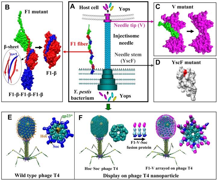
Figure 1. New plague immunogen designs.
Schematic of various approaches used to design plague immunogens. See text for details. (A) Y. pestis surface components targeted for vaccine design. F1 is the structural unit of the capsular layer. V forms a pore at the tip of the injectisome needle and facilitates translocation of Yops into the host cell. YscF is the structural unit of the injectisome needle. (B) Reorientation of the NH2-terminal β-strand of F1 to generate monomeric F1. “n” and “n+1” refer to the F1 subunits the β-strands belong to; the red strands to “n” subunit and the blue strand to the “n+1” subunit. (C) Deletion of the putative immunomodulatory sequence (aa residues 271–300) of V antigen. (D) Mutagenesis of Asn35 and Ile67 to produce an oligomerization deficient YscF. (E) Structural model of bacteriophage T4. The enlarged capsomer shows the major capsid protein gp23* (green; “*” represents the cleaved form) (930 copies), Soc (blue; 870 copies), and Hoc (yellow; 155 copies). Yellow subunits at the five-fold vertices correspond to gp24*. The portal vertex (not visible in the picture) connects the head to the tail. (F) Display of F1mut-V-Soc fusion protein on the Hoc− Soc− phage particle. Models of the enlarged capsomers before and after F1mut-V display are shown.

