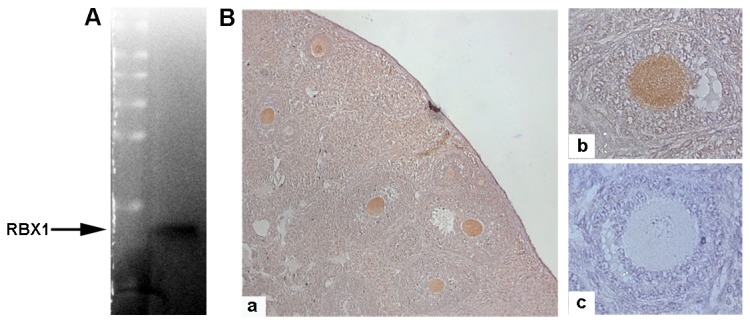
Figure 1. Presence of RBX1 in mouse oocytes.

A) Western blot image showing the only band with the predicted molecular weight of 14 kD, indicating the presence of RBX1 in mouse oocytes. B) Immunohistochemistry images showing the abundant presence of RBX1 in oocytes (a. 10×, b. 40×, c. negative control).
