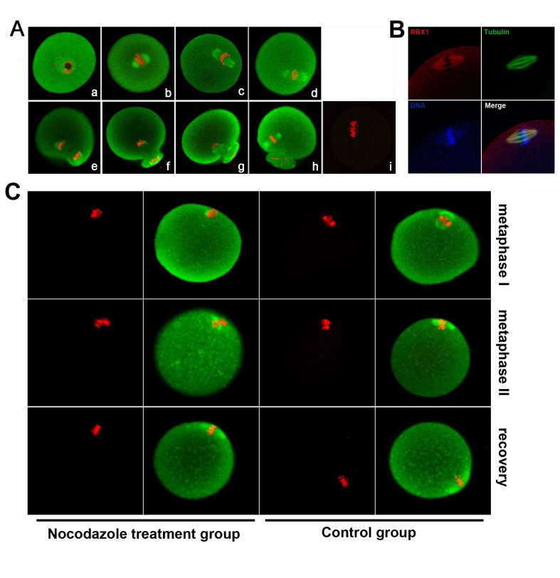
Figure 2. Subcellular localization of RBX1 during mouse oocyte meiotic maturation.
A) Confocal microscope image showing immunostaining of RBX1 (green) and DNA (red) in oocytes at GV (a), GVBD+2 h (b), GVBD+4 h (c), GVBD+6 h (d), GVBD+8 h (e), GVBD+10 h(f), GVBD+12 h (g), and GVBD+16 h (meiosis II stage) (h), rabbit IgG was used as a negative control (i). B) Image of in vivo mature eggs double-stained with antibodies against RBX1 and α-tubulin: green, α-tubulin; red, RBX1; blue, chromatin; yellow, RBX1 and α-tubulin overlapping. C) Images of oocytes treated with nocodazole for spindle perturbation and then treated with fresh medium for spindle recovery. Oocytes at metaphase I and metaphase II stage were treated with nocodazole for spindle perturbation, and then MII oocytes were transferred to fresh CZB after washing away nocodazole for spindle recovery. Green, RBX1; red, DNA.

