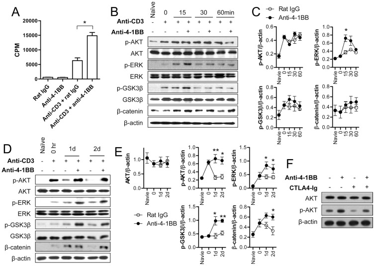
Figure 1. 4-1BB sustains anti-CD3 induced signals to induce GSK-3/β-catenin signaling with delayed kinetics.
The purified CD8+ T cells from C57BL/6 mice were activated with 0.1 µg/ml of anti-CD3 for 16 h. (A) CD8+ T cells cultured with or without anti-CD3 for 16 h were further incubated with 5 µg/ml of anti-4-1BB or rat IgG for another 48 h, and cell proliferation was assessed by [3H]-thymidine incorporation. (B–C) The activated CD8+ T cells were incubated with 5 µg/ml of anti-4-1BB or rat IgG for 0, 15, 30, or 60 min. Western blot analysis was performed with p-AKT, p-ERK, p-GSK-3β, β-catenin, and β-actin antibodies (B) and the relative expression levels of p-AKT, p-ERK, p-GSK-3β, and β-catenin were determined (C). (D–E) The activated CD8+ T cells were stimulated with 5 µg/ml of anti-4-1BB for 0, 1, or 2 days. Western blotting was performed (D) and the relative expression levels of each protein was determined (E). (F) The activated CD8+ T cells were treated with 5 µg/ml anti-4-1BB or rat IgG following 1 h incubation with 10 µg/ml CTLA-4Ig. After 24 h incubation, CD8+ T cells were harvested, lysed and subjected to Western blot analysis using AKT, p-AKT, and β-catenin antibodies. Naïve indicates CD8+ T cells that were cultured in complete medium for 16 h without anti-CD3. Results in C and E are mean ±SD of three independent experiments and representative data of two independent experiments are shown in F (*p<0.05; **p<0.01).

