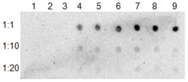
Figure 4. Surface localization in E. coli of GFP fusion proteins of LasAI and LasAII by immuno-dot blot.

Serial dilutions of bacterial cells were deposited onto a nitrocellulose membrane; GFP was detected with anti-GFP antibody. Lane 1, 2, 3: IPTG induced E. coli containing plasmid pET102-gfp; Lane 4, 5, 6: IPTG-induced E. coli containing plasmid pET102-gfp-lasA I-TD; Lane 7, 8, 9: IPTG induced E. coli containing plasmid pET102-gfp-lasA II.
