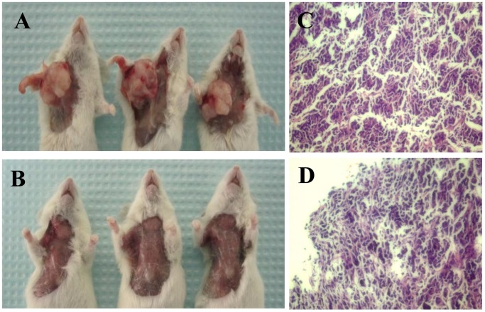
Figure 5. Tumor formation of sorted 769P SP and NSP cells in NOD/SCID mice and pathological examination with H&E staining.
A and B, injection sites of NOD/SCID mice at 6 weeks after inoculation with freshly sorted SP or NSP cells. Tumors are observed in all mice inoculated with 2,000 SP cells, but not in mice inoculated with 2,000 NSP cells. C and D, Pathological examination shows typical renal cell carcinoma in mice inoculated with either 2,000 SP cells or 200,000 NSP cells.

