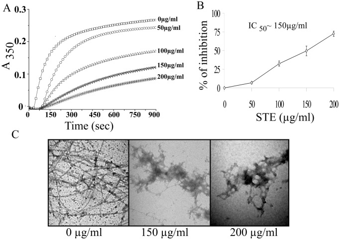
Figure 7. Inhibition of the assembly of purified tubulin by STE.
(A) Tubulin assembly study. Tubulin (12 µM) was polymerized separately in the presence of (0 −200 µg/ml) STE at 37°C. The progress of tubulin assembly was monitored spectrophotometrically at 350 nm. (B) A plot of percentage of polymerization inhibition against dose of STE. Data represent the mean ±SEM (p<0.05 vs control, n = 3). (C) Aggregation of microtubule protofilaments in the presence of STE as observed by a transmission electron microscopy. Tubulin (12 µM) was polymerized separately in the presence of different STE doses (0–200 µg/ml), and the images were taken at 20000X magnification. The bar represents 500 nm. The results represent the best of data collected from three experiments with similar results.

