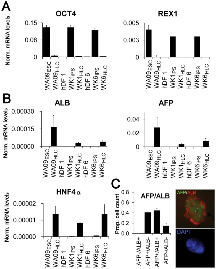
Figure 4. Expression of pluripotency and definitive hepatocyte markers before and after iPS, and in HLCs derived from pluripotent cells.
(A) Analysis of expression of the pluripotency markers OCT4 and REX1 by qRT-PCR in: undifferentiated hESCs (WA09ESC) and stage 3B HLCs derived from WAO9 (WAO9HLC); dermal fibroblast line hDF1, hiPSC line WK1 derived from hDF1 (WK1iPS) and stage 3B hepatocyte-like cells derived from WK1 (WK1HLC); and dermal fibroblast line hDF6, the hiPSC line WK6 derived from hDF1 (WK1iPS) and stage 3B hepatocyte-like cells derived from WK6 (WK6HLC). (B) Induction of expression of the hepatocyte markers ALB, AFP, and HNF4α by qRT-PCR before and after reprogramming, and after stage 3B differentiation (for cell-type nomenclature see Fig. 3A legend). (C) Cell counts for AFP and ALB expression in cytocentrifuged stage 3B cells. WK1HLCs were labeled with anti-human ALB and anti-human AFP antibodies and quantified as described (see Experimental Procedures). The inset depicts a representative high magnification image showing AFP (green) and ALB (red) expression in the top panel and DAPI (blue) in the bottom panel. Error bars represent the standard error of the mean.

