Figure 4.

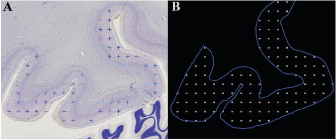
Illustration of the data acquisition in Stereo Investigator. A. Image of the Nissl-stained FG with markers indicating counted neurons; B. The outline of the sampled ROI and the grid of acquired image stacks in which neurons were counted.

Illustration of the data acquisition in Stereo Investigator. A. Image of the Nissl-stained FG with markers indicating counted neurons; B. The outline of the sampled ROI and the grid of acquired image stacks in which neurons were counted.