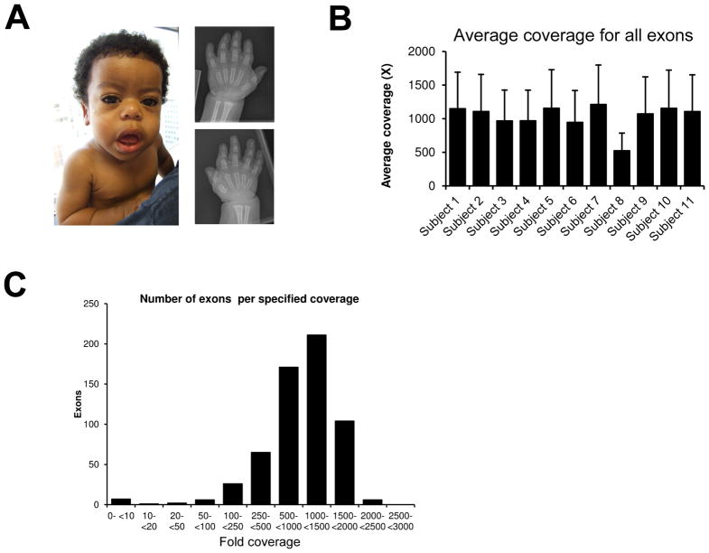
Figure 1.
A) Subject 6 with a diagnosis of geleophysic dysplasia. Photograph showing classical clinical features and hand radiograph demonstrating generalized shortening of phalanges and metacarpals. B) The average coverage per patient for all exons was above 400X. C) Distribution of coverage per exon by the NGS panel.

