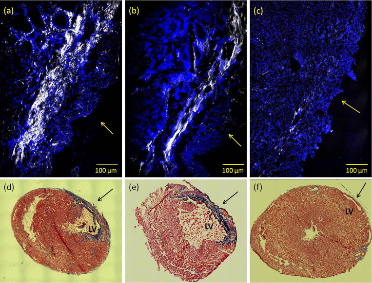
Figure 1. Co-localized SHG (shown in white, un-labeled) and TPEF (shown in blue, labeled with DAPI) images visualize collagen fibril organization and cardiac muscle cell nuclei, respectively, in the histological section of infarcted myocardium of (a) an untreated infarcted rat heart; (b) an ASCs-treated infarcted rat heart; (c) an image obtained from a histological section of a non-MI rat heart.
Images were acquired using 10× 0.45NA dry objective lens. Excitation wavelength is at 800 nm. Collagen SHG signal was collected using a 400 ± 5 nm band-pass filter in the forward direction while the DAPI -TPEF signal was collected in the backscattered (epi) direction through a 505 ± 50 nm filter. Arrows are pointing to the epicardium region. (d), (e) and (f) show representative short-axis histopathological sections of untreated, ASCs-treated infarct rat heart and non-MI heart, respectively. Heart tissue sections were stained with Masson's Trichrome to delineate the infarct region, and images were acquired using 5× objective lens. LV: left-ventricle.

