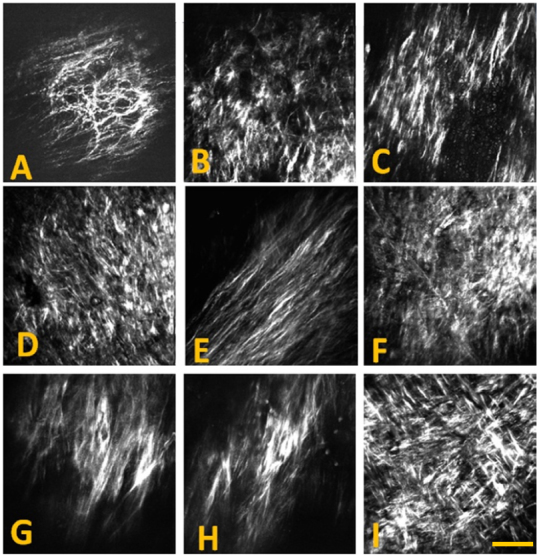
Figure 4. Epi-SHG images acquired from atherosclerotic plaques on the aorta of WHHL rabbits, showing examples of different collagen fibril morphology detected on atherosclerotic artery.

SHG images were acquired using 20 × 0.75 NA dry objective lens (Olympus) and 800 nm laser excitation. A 2× digital zoom was used for imaging. Each image shown has 512 × 512 pixels or approx. 200 × 200 μm. Scale bar: 50 μm.
