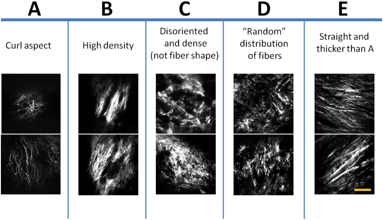
Figure 5. All collagen SHG images acquired from the arteriosclerotic aortic segments of the WHHL rabbits were classified into five groups A–E.
Each group of the images has its own characteristic morphological features such as the fibril's shape, size and organization. Images are showing the fibrous cap, accumulated closer to the intima layer. Representative images from each group (A–E) are shown. Scale bar: 50 μm.

