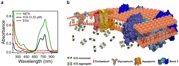
Figure 2. Absorption spectra and a physical model of a NET containing ICG.
(a), Absorption spectra corresponding to free (non-encapsulated) ICG (3.22 μM) dissolved in PBS, and EGs and NETs re-suspended in PBS after fabrication. (b), A physical model of a NET showing an ensemble of ICG conformational states comprised of ICG monomers, ICG aggregates, and monomers and aggregates of ICG bound to membrane lipids and/or membrane proteins. For illustration purposes, we present three main membrane integral proteins of erythrocytes: Aquaporin, Band3 and Glycophorin.

