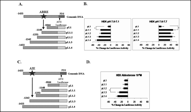
Fig. 2.
Effect of ambient pH (A and B) and of aldosterone (C and D) on the hPDS promoter in HEK293 cells. A and C: schematic representation of the DNA constructs of hPDS promoter in the luciferase reporter vector, pGL3-basic, used in these experiments. ABRE, putative acid-base response element; AIE, putative aldosterone-induced element. B and D: cells were transfected with pGL3-basic or pGL3-basic containing segments of decreasing size corresponding to the 5'-flanking region of hPDS (pL1.4, pL1.3, pL1.2, pL1.1, and pL1). Cells were exposed to acidic pH (7.0-7.1), normal pH (7.35-7.45), or alkaline pH (7.6-7.7) (B) or to 10-8 M aldosterone (D) for 24 h. Subsequently, luciferase activity was measured. To control for transfection efficiency, cells were cotransfected with the LacZ- containing vector, pCH110, and luciferase activity was normalized to β-galactosidase activity. Data represent the %change in luciferase activity in cells exposed to experimental media (with acidic pH, alkaline pH, or aldosterone) relative to cells exposed to control medium (pH 7.35-7.45 without aldosterone). Values are means ±SE of 3-5 independent experiments, each performed in quadruplicate. Acidic pH-induced inhibition and alkaline pH-induced stimulation of luciferase activity were evident in HEK293 cells, which markedly decreased when the fragment size was shortened from 1.1 kb to 1 kb (B). Aldosterone-induced inhibition of luciferase activity was demonstrated in HEK293 cells, which markedly diminished when the fragment size was truncated from 1.3 kb to 1.2 kb (D). *P < 0.01 (modified from reference [42]).

