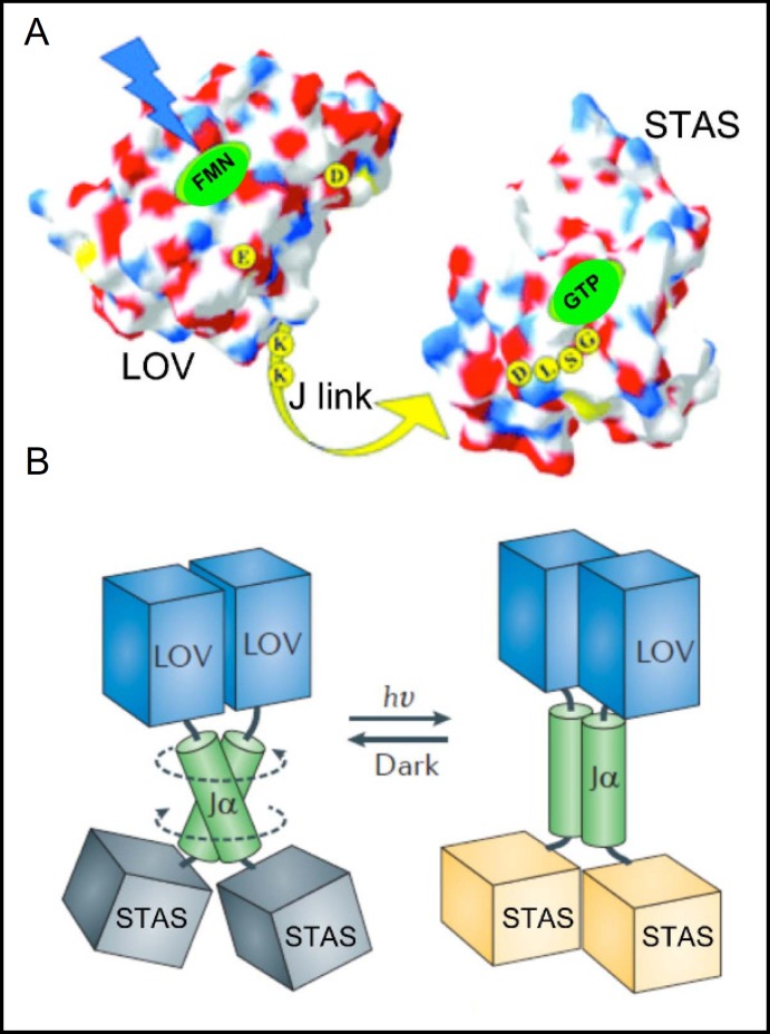
Fig. 3.
A. B. subtilis YtvA electrostatic surface image from LOV domain crystal structure and from STAS domain (modeled on crystal structure of B. subtilis SpoIIAA). Blue light is absorbed by the flavin mononucleotide chromophore of the LOV domain, triggering local conformational change that is believed to be transmitted through the agency of the J linker to the STAS domain, which binds BODIPY-GTP. Yellow residues have been implicated by mutagenesis as important or required for phototransduction. Modified from [21]. B. Light-induced conformational change of YtvA as imagined from holoprotein structure. Modified from [26].

