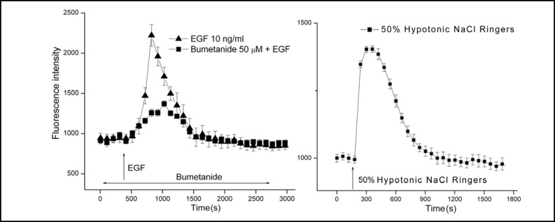
Fig. 2.
EGF induces transient HCEC swelling through NKCC1 activation. Calcein loaded HCEC attached to coverslips were placed in a chamber for superfusion of room temperature NaCl Ringers from syringes onto the stage of an epifluorescent microscope. The epifluorescent emission was measured between 503 and 530 nm. Panel A shows that following signal stabilization of about 3 min during control superfusion, the solution was substituted for another containing 10 ng/ml EGF (T) (Mean ± SEM: n=5 coverslips; p<0.001 versus untreated control). After about 3 min, the corrected signal nearly rapidly doubled followed by gradual decline to reach a baseline level after about 15 more min. Pre-exposure for 30 min to 50 μM bumetanide suppressed the subsequent EGF-induced transient by about 70% (n=5; p<0.001). Panel B shows that substitution of the control 306 mOsm NaCl Ringers with one diluted by 50% induced after about 1 min a transient increase in fluorescence reflective of swelling since calcein is a self-quenching dye (n=4 coverslips; p<0.005 versus isotonic control). The pattern of this transient was very similar to that induced by EGF, but instead is reflective of hypotonic-induced stimulation of KCC and K+ channel activity rather than NKCC1 activation by EGF shown in panel A [7, 17].

